Home
/ Plant Cell As Seen Under A Light Microscope - Microscopy How A Microscope Works Magnification Calculations How To Use A Microscope Slide Preparation Investigations Resolution Resolving Power Measuring Size Of Cell Electron Microscope Micrograph Light Micrograph Igcse O Level Gcse 9 1 Biology Revision - The animal cell is more fluid or elastic or malleable in structure;
Plant Cell As Seen Under A Light Microscope - Microscopy How A Microscope Works Magnification Calculations How To Use A Microscope Slide Preparation Investigations Resolution Resolving Power Measuring Size Of Cell Electron Microscope Micrograph Light Micrograph Igcse O Level Gcse 9 1 Biology Revision - The animal cell is more fluid or elastic or malleable in structure;
Plant Cell As Seen Under A Light Microscope - Microscopy How A Microscope Works Magnification Calculations How To Use A Microscope Slide Preparation Investigations Resolution Resolving Power Measuring Size Of Cell Electron Microscope Micrograph Light Micrograph Igcse O Level Gcse 9 1 Biology Revision - The animal cell is more fluid or elastic or malleable in structure;. Below is a size and. Cells of plant or animal tissue. Plant cell organelles that are invisible under a compound light microscope include mitochondria, ribosomes, endoplasmic reticula, and golgi bodies. The animal cell is more fluid or elastic or malleable in structure; Light microscopes also have a shallow depth of field.
Living multicellular organisms of the kingdom plantae. Below is a size and. The easiest and least complicated solution to view bacteria using light microscopes is to buy a prepared permanent slide. Cells are generally microscopic, so you need a microscope to see the parts they are made of. Beneath a plant cell's cell wall is a cell membrane.
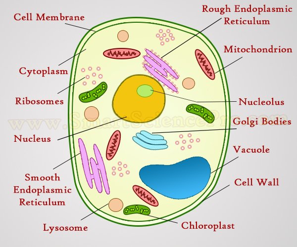
Structure Of Animal Cell And Plant Cell Under Microscope Diagrams from www.smartsciencepro.com This is a single cheek cell that has been stained, as seen under a light microscope. Made of cellulose and gives the cell structure. (ii) presence of large central vacuole in plant cell. Living multicellular organisms of the kingdom plantae. (iii) presence of cell how its different from animal cell? Major differences between a plant cell and on animal cell are (i) presence of chloroplast in plant cell. Plant cell organelles that are invisible under a compound light microscope include mitochondria, ribosomes, endoplasmic reticula, and golgi bodies. Image:animal cell seen under light microscope.
You observe plant cells under a microscope as they are placed in an unknown solution.
The plant cell as more rigid and stiff walls. Draw a neat diagram of plant cell and label any three parts which differentiate it from animal cell. Step by step solution by experts to help you in doubt clearance & scoring excellent marks in exams. A microscope allows you to see detail in specimens that you cannot see. The animal cell is more fluid or elastic or malleable in structure; An electron microscope is required for virus and dna. They were compiled by ge healthcare these images, compiled by ge healthcare life sciences as part of an annual competition, reveal the beautiful views scientists see as they peer into. Under a light microscope, the cell membrane, nucleus and cytoplasm of a cheek cell (animal cell) can be observed. Thus, we need the amplifying power of the microscope to see cells and even the structure and organelles inside of cells. Made of cellulose and gives the cell structure. Answer the following questions in your exercise book. Cell structures as seen using a light microscope. Cells of plant or animal tissue.
You see that many features are in common. .between a plant cell and on animal cell are (i) presence of chloroplast in plant cell. Under a microscope, plant cells from the same source will have a uniform size and shape. Plant cells have an additional layer surrounding the cell on the outside of the cell membrane. Light microscopy is the most accessible form of microscopy around the world and.
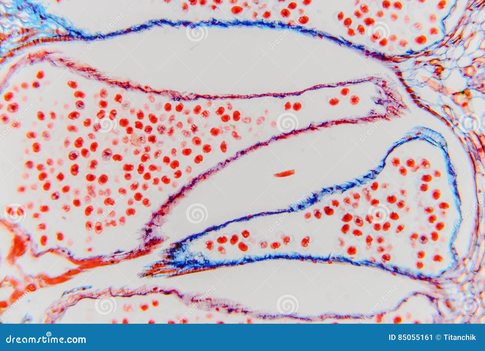
Plant Cell High Magnification With Light Microscope Science Ba Stock Image Image Of Leaf Botanical 85055161 from thumbs.dreamstime.com Cells are generally microscopic, so you need a microscope to see the parts they are made of. Plant cells have an additional layer surrounding the cell on the outside of the cell membrane. The animal cell is more fluid or elastic or malleable in structure; But in animals, there is a cell membrane n. See how a generalized structure of an animal cell and plant cell look with labeled diagrams. Plant cell organelles that are invisible under a compound light microscope include mitochondria, ribosomes, endoplasmic reticula, and golgi bodies. (iii) presence of cell wall. Living multicellular organisms of the kingdom plantae.
Click (or tap) the diagram for a simple labelled version.
While observing plant cells in a leaf with my students, i noticed small black particles moving erratically about in some of the. Light microscopes also have a shallow depth of field. (ii) presence of large central vacuole in plant cell. Animal and plant cells have features in common and differences between animal. What cell organelles can be seen under the electron microscope but not with the light microscope and their functions in the cell? This is a single cheek cell that has been stained, as seen under a light microscope. A cell is a very tiny structure which exists in living bodies. In this lab reportt you will see how the cells are seen under the microscopes and will know how to introduction compound light microscope: Animal cells under a light microscope. Cells are generally microscopic, so you need a microscope to see the parts they are made of. Most cells are visible under a light microscope, but mitochondria and bacteria are barely visible. It also has a very high resolving power. But in animals, there is a cell membrane n.
Cell is a tiny structure and functional unit of a living organism containing various parts known as organelles. Is all the living parts of a cell. Most cells are visible under a light microscope, but mitochondria and bacteria are barely visible. This gives rise to its name of rough endoplasmic reticulum (often structures in a plant cell visible under a light microscope and an electron microscope. Microscope capable of increasing our ability to see by plant cell:
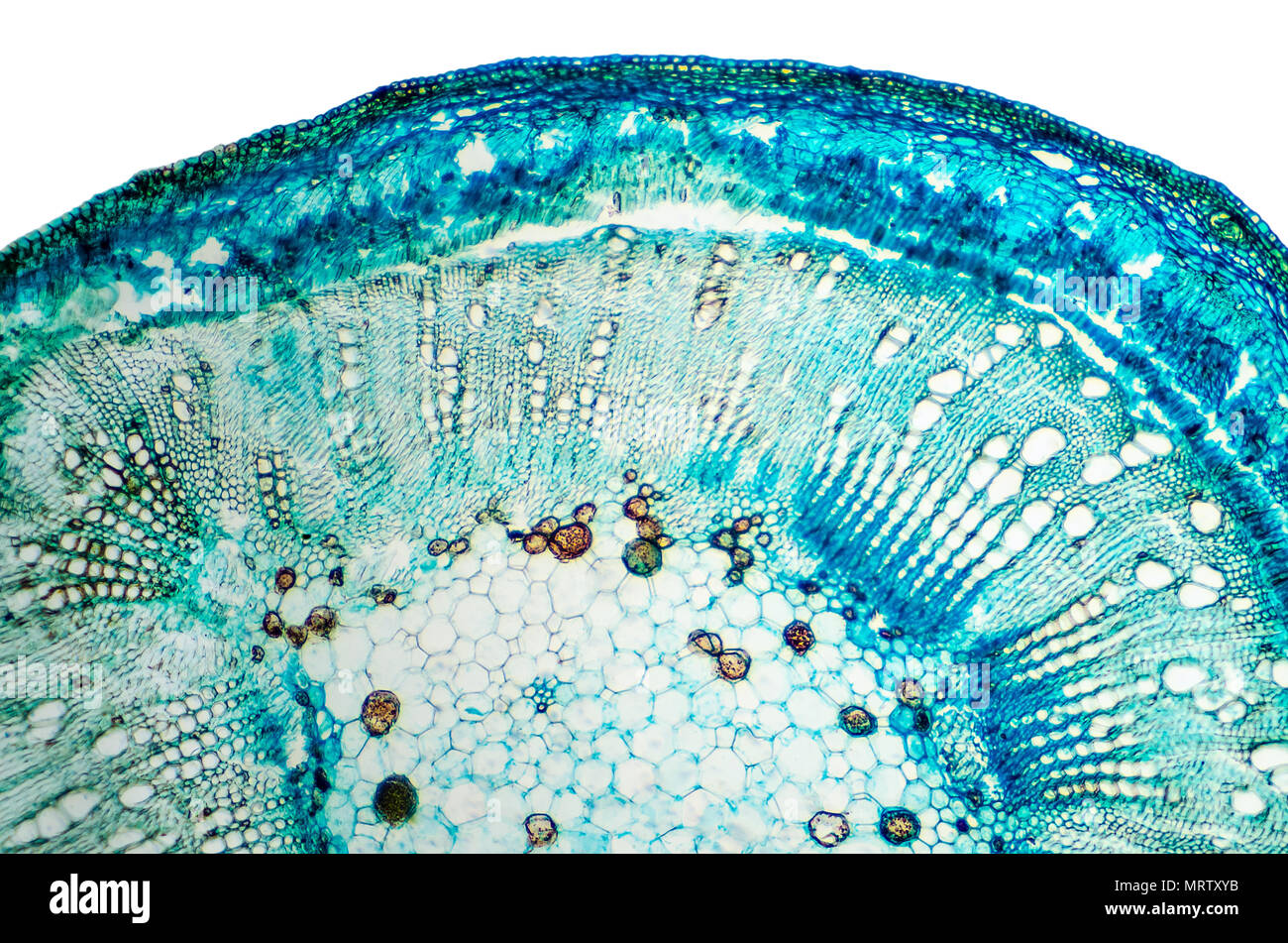
Plant Cells Microscope High Resolution Stock Photography And Images Alamy from c8.alamy.com Describe and compare the structure of a plant cell with an animal cell, as seen under a light microscope, limited to cell wall, nucleus, cytoplasm, chloroplasts, vacuoles and location of the cell membrane. Resolving power is the ability to distinguish between separate things which are close to each other. A scale bar has been marked on the drawing, allowing the size of a cell to be estimated. The scientists schleiden and schwann observed plant and animal tissues under the microscope. Light microscopes also have a shallow depth of field. Plant cells have an additional layer surrounding the cell on the outside of the cell membrane. If you take some pollen, and sprinkle a little (the examine your preparation under a 10x and a 40x objective and observe the starch grains. This gives rise to its name of rough endoplasmic reticulum (often structures in a plant cell visible under a light microscope and an electron microscope.
A scale bar has been marked on the drawing, allowing the size of a cell to be estimated.
This is a single cheek cell that has been stained, as seen under a light microscope. Draw the outline of a few starch grains as seen under hp (at. An electron microscope is required for virus and dna. Studying tissue with light microscopy has been practiced for more than 50 years. Endoplasmic reticulum studded with ribosomes looks rough under the microscope; Light microscopes also have a shallow depth of field. .between a plant cell and on animal cell are (i) presence of chloroplast in plant cell. Draw a neat diagram of plant cell and label any three parts which differentiate it from animal cell. They were compiled by ge healthcare these images, compiled by ge healthcare life sciences as part of an annual competition, reveal the beautiful views scientists see as they peer into. Animal and plant cells have features in common and differences between animal. Describe and compare the structure of a plant cell with an animal cell, as seen under a light microscope, limited to cell wall, nucleus, cytoplasm, chloroplasts, vacuoles and location of the cell membrane. Made of cellulose and gives the cell structure. But in animals, there is a cell membrane n.
between a plant cell and on animal cell are (i) presence of chloroplast in plant cell plant cell under a light microscope. The animal cell is more fluid or elastic or malleable in structure;
Share :
Post a Comment
for "Plant Cell As Seen Under A Light Microscope - Microscopy How A Microscope Works Magnification Calculations How To Use A Microscope Slide Preparation Investigations Resolution Resolving Power Measuring Size Of Cell Electron Microscope Micrograph Light Micrograph Igcse O Level Gcse 9 1 Biology Revision - The animal cell is more fluid or elastic or malleable in structure;"

Post a Comment for "Plant Cell As Seen Under A Light Microscope - Microscopy How A Microscope Works Magnification Calculations How To Use A Microscope Slide Preparation Investigations Resolution Resolving Power Measuring Size Of Cell Electron Microscope Micrograph Light Micrograph Igcse O Level Gcse 9 1 Biology Revision - The animal cell is more fluid or elastic or malleable in structure;"